Taping technique for medial epicondylitis or golfer’s elbow
This is an excerpt from A Practical Guide to Kinesiology Taping for Injury Prevention and Common Medical Conditions-3rd Edition by John Gibbons.
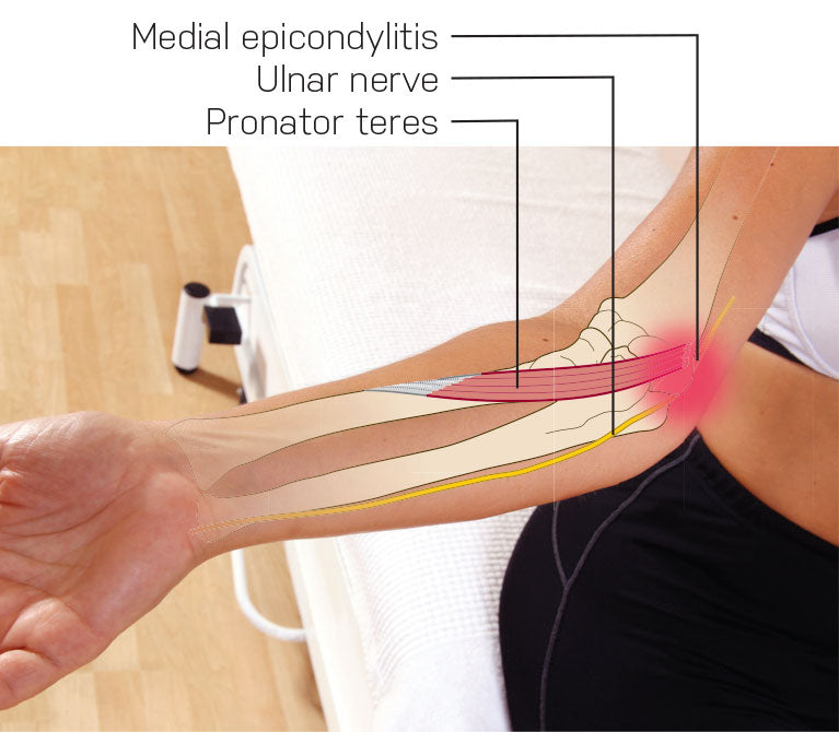
When I was a physical trainer with the army I liked climbing ropes as I found this exercise very effective for building my strength. However, over time I would feel pain to the inside aspect of my elbow and I was told I had golfer’s elbow, even though I did not play golf. This condition can basically affect anybody and it is normally caused by one particular muscle, pronator teres (Fig. 8.5). This muscle originates at the medial epicondyle; hence, the term ‘medial epicondylitis’. There is also a relationship to the ulnar nerve due to the proximity of the inflammation. If a patient perceives a tingling or altered sensation to their little finger then the ulnar nerve is also involved.

The following kinesiology taping technique will help both the pronator teres and the ulnar nerve.
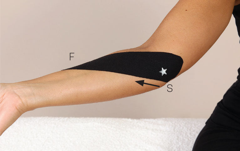
- Place the pronator teres muscle into a stretched position by supinating the forearm and extending the elbow. Then apply an ‘I’ strip, with little to no stretch, starting from the origin point on the medial epicondyle and finishing across the pronator teres (Fig. 8.6).

Figure 8.6 First application of an ‘I’ strip to the pronator teres.
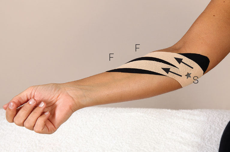
- Apply a smaller than standard ‘Y’ strip, with 75–100% stretch, across the area of pain (Fig. 8.7).

Figure 8.7 Second application of a smaller ‘Y’ strip across the area of pain.
- Heat activate the glue.
SHOP

Get the latest insights with regular newsletters, plus periodic product information and special insider offers.
JOIN NOW
Latest Posts
- Women in sport and sport marketing
- Sport’s role in the climate crisis
- What international competencies do sport managers need?
- Using artificial intelligence in athletic training
- Using the evidence pyramid to assess athletic training research
- How can athletic trainers ask a clinically relevant question using PICO?


