Transmission of a nerve impulse
This is an excerpt from The Pocket Atlas of Anatomy and Physiology by Ruth Hull.
Nerve impulses are transmitted electrochemically either across the plasma membrane of an unmyelinated axon or across the nodes of Ranvier of a myelinated axon.
A nerve impulse is generated and propagated as follows:
An inactive plasma membrane has a resting membrane potential that is polarized. This means that there is an electrical voltage difference across the membrane, with the external voltage being positive and the internal one being negative. The main external ions are sodium, while the main internal ones are potassium.
When the dendrites of the neuron are stimulated, ion channels in a small segment of the plasma membrane open and allow the movement of sodium ions into the cell. This causes the inside of the cell to become positive and the outside negative. This is called “depolarization.”
Depolarization causes the membrane potential to be reversed and this initiates an action potential (impulse). When one segment of the membrane becomes depolarized, it causes the segment next to it to be depolarized, and so a wave of depolarization is propagated down the length of the plasma membrane. This is how the action potential travels to the end of the neuron.
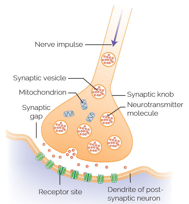
When the action potential reaches the end of the neuron, it causes vesicles containing neurotransmitters to open up and release a neurotransmitter into the synaptic cleft. The neurotransmitter diffuses across the synaptic cleft and binds to the receptors of the next neuron, muscle, or gland, where it now acts as a stimulus.

Nerves
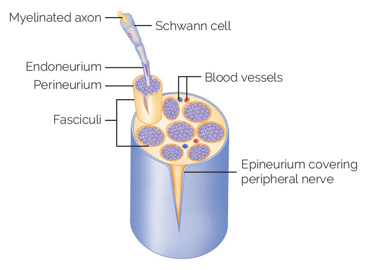
A nerve consists of a bundle of nerve fibers surrounded by connective tissue.


SHOP

Get the latest insights with regular newsletters, plus periodic product information and special insider offers.
JOIN NOW


